Bone Cross Section Diagram / If You Have Osteosarcoma / Explaned distal and proximal epiphysis.. Two prominent grooves or sulci run along its length. Cross sections are 2d educated guesses at the geology along a plane different from the surface of the earth. In a cross section of a bone we can see two types of bone tissue: I am not an expert on this subject, so i was wondering if anyone could put their input on it seems confusing and misleading. From wikimedia commons, the free media repository.
Diagram with articular cartilage, marrow, spongy bone, medullary cavity, endosteum, diaphysis, and periosteum. So what im going to do is jump right to the. Diagram with articular cartilage, marrow, spongy bone, medullary cavity, endosteum, diaphysis, and periosteum. Explaned distal and proximal epiphysis. Vector illustration scheme of bone cross section.
Adaptation of bone geometry. (a) Cross-section of long ... from www.researchgate.net Bone is found in the shafts of long bone and consists of various cylindrical units named as haversian system 47. Diagram with articular cartilage, marrow, spongy bone, medullary cavity, endosteum, diaphysis, and periosteum. Tetraplegia and paraplegia spinal neural disorder medical vector illustration diagram with female back bone cross section. Spongy bone and compact bone. Vector illustration scheme of bone cross section. Explaned distal and proximal epiphysis. A cross section diagram is if you would take a knife and cut through one side of a diagram to see the inside and outside in one picture. Hi all, i have uploaded a new medical animation tutorial.
A cross section of a human long bone.
Crosssection cutaway diagram dry cell battery. Histology sauropod vertebra picture of the week these pictures of this page are about:long bone cross section. Cross section diagrams are used a lot by architects and engineers to show what a building or machine might look like before it's built. From wikimedia commons, the free media repository. Diagram with articular cartilage, marrow, spongy bone, medullary cavity, endosteum, diaphysis, and periosteum. Ear external and internal anatomy cross section unlabeled stock illustration 9895a hr fotosearch / wh. The centroidal locations of common cross sections are well documented, so it is typically not necessary to calculate the location with the equations above. Cross section of a bone diagram : For example, to read this diagram literally, since the cartilage can be seen inside the cutaway section of bone, it. Explaned distal and proximal epiphysis. Cross sections are 2d educated guesses at the geology along a plane different from the surface of the earth. The last problem asks you to balance a crosssection, and. Vector illustration scheme of bone cross section.
Hi all, i have uploaded a new medical animation tutorial. Diagram with articular cartilage, marrow, spongy bone, medullary cavity, endosteum, diaphysis, and periosteum. Diagram with articular cartilage, marrow, spongy bone, medullary cavity, endosteum, diaphysis, and periosteum. Diagram with articular cartilage, marrow, spongy bone, medullary cavity, endosteum, diaphysis, and periosteum. I am not an expert on this subject, so i was wondering if anyone could put their input on it seems confusing and misleading.
Cross-section of hip joint to reveal internal bone ... from fscomps.fotosearch.com As with other tools applied to petroleum development. As with other tools applied to petroleum development. Explaned distal and proximal epiphysis. Tetraplegia and paraplegia spinal neural disorder medical vector illustration diagram with female back bone cross section. Vector illustration scheme of bone cross section. The centroidal locations of common cross sections are well documented, so it is typically not necessary to calculate the location with the equations above. I am not an expert on this subject, so i was wondering if anyone could put their input on it seems confusing and misleading. Ear external and internal anatomy cross section unlabeled stock illustration 9895a hr fotosearch / wh.
The centroidal locations of common cross sections are well documented, so it is typically not necessary to calculate the location with the equations above.
Explaned distal and proximal epiphysis. At present, however, it seems this remains difficult to. This article covers the anatomy of the spinal cord including its structure tracts and function. As with other tools applied to petroleum development. Two prominent grooves or sulci run along its length. Each system contains haversian canals surrounded by concentric lamellae of bone tissue 48. For example, to read this diagram literally, since the cartilage can be seen inside the cutaway section of bone, it. In this short video i use blender 2.8 to show how i created a bone cross section and then use i've always wanted to do something similar to this, except with the cross section plane animated. Histology sauropod vertebra picture of the week these pictures of this page are about:long bone cross section. Explaned distal and proximal epiphysis. In a cross section of a bone we can see two types of bone tissue: I am not an expert on this subject, so i was wondering if anyone could put their input on it seems confusing and misleading. The centroidal locations of common cross sections are well documented, so it is typically not necessary to calculate the location with the equations above.
Vector illustration scheme of bone cross section. At present, however, it seems this remains difficult to. Diagram with articular cartilage, marrow, spongy bone, medullary cavity, endosteum, diaphysis, and periosteum. Editable vector illustrator cc file (editable live text)editable vector eps 10 filehigh. Hi all, i have uploaded a new medical animation tutorial.
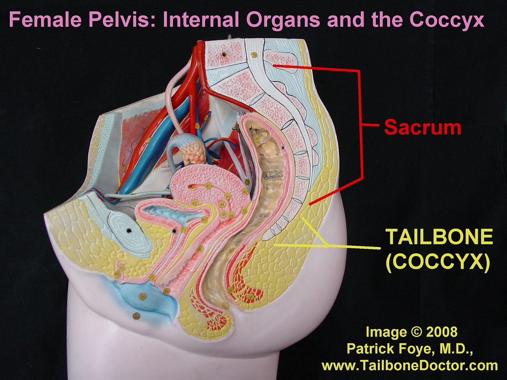
Female Pelvis, Tailbone, Coccyx | This is a cross section ... from c2.staticflickr.com Diagram with articular cartilage, marrow, spongy bone, medullary cavity, endosteum, diaphysis, and periosteum. Most relevant best selling latest uploads. Explaned distal and proximal epiphysis. Explaned distal and proximal epiphysis. Cross section of a bone diagram : A cross section diagram is if you would take a knife and cut through one side of a diagram to see the inside and outside in one picture. Tetraplegia and paraplegia spinal neural disorder medical vector illustration diagram with female back bone cross section. Although this plane is almost always vertical this lab is designed introduce you to the basic techniques of building cross sections.
Editable vector illustrator cc file (editable live text)editable vector eps 10 filehigh.
Jump to navigation jump to search. Diagram with articular cartilage, marrow, spongy bone, medullary cavity, endosteum, diaphysis, and periosteum. At present, however, it seems this remains difficult to. So what im going to do is jump right to the. Spongy bone and compact bone. Brain cross section diagram illustrations & vectors. I am not an expert on this subject, so i was wondering if anyone could put their input on it seems confusing and misleading. (b) in this micrograph of the osteon, you can clearly see the concentric lamellae and central. As with other tools applied to petroleum development. Histology sauropod vertebra picture of the week these pictures of this page are about:long bone cross section. A cross section of a human long bone. This is a short tutorial using blender 2.8 that shows how to create a bone cross section and using images to create the textures.hope you enjoy and please. Explaned distal and proximal epiphysis.
The last problem asks you to balance a crosssection, and bone cross section. So what im going to do is jump right to the.


0 Komentar